
For patient education: Maneuver photographs are conceptual illustrations, not validated self-treatment instructions. Correct canal, side and subtype selection requires clinical assessment; neck/vascular conditions and fall risk must be considered. Do not attempt these maneuvers without appropriate professional guidance.
Why the room may spin when you roll over, how clinicians identify the affected ear, and which repositioning maneuvers may help.
Have you ever rolled over in bed, tilted your head back, or bent down and suddenly felt the room spinning? If the sensation comes in brief episodes triggered by particular head positions, one possible cause is benign paroxysmal positional vertigo (BPPV), a common inner-ear disorder.
BPPV can cause intense spinning, nausea, and unsteadiness. The encouraging news is that many cases respond well to precise, clinician-guided repositioning maneuvers. The important first step is determining whether BPPV is actually the cause—and, if it is, which ear and semicircular canal are involved.

What are the “crystals” in the inner ear?
Inside each inner ear are three fluid-filled semicircular canals—posterior, horizontal (lateral), and anterior (superior)—that help detect head rotation. Nearby, the utricle senses head tilt and linear movement. The utricle contains tiny calcium-carbonate particles called otoconia, normally embedded in a gelatinous layer above sensory hair cells.
When otoconia become displaced into a semicircular canal, head movements may shift the particles and disturb the normal sensory signal. The brain can then receive a false impression of rotation, producing a sudden spinning sensation. BPPV can occur without a clear trigger, but it may follow head injury or become more common with age.
Typical BPPV: brief spinning triggered by looking up, bending over, lying back, or rolling in bed. Nausea and temporary imbalance may accompany an episode. Persistent dizziness, fainting, neurological symptoms, or hearing changes require consideration of other causes.
How clinicians diagnose BPPV—and identify the right or left ear
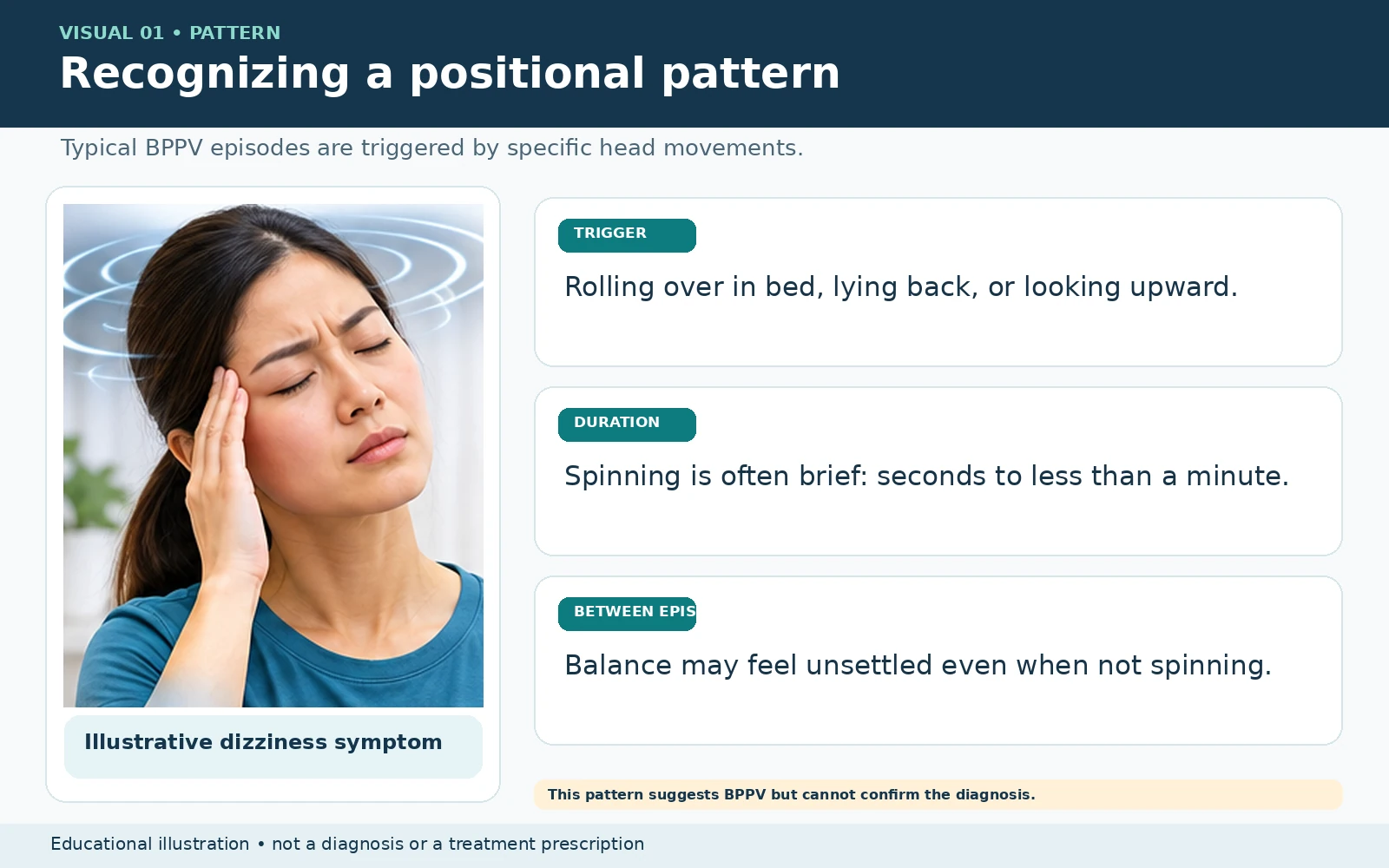
A clinician first reviews the pattern of dizziness, checks for warning signs, and considers neck mobility and other conditions that could affect testing. During positional tests, the clinician looks for nystagmus—a characteristic pattern of involuntary eye movement. Its direction and relative strength can help identify the affected canal and side.
| Suspected location | Main assessment | Typical finding and right/left clue | Common treatment |
|---|---|---|---|
| Posterior canal Most common | Dix–Hallpike test on each side, when safe | Brief upbeating, torsional nystagmus. A characteristic response with the right ear down points to right posterior-canal BPPV; a corresponding left-ear-down response points to the left. | Epley or Semont maneuver, matched to the affected side. |
| Horizontal canal | Supine roll test to the right and left | Direction-changing horizontal nystagmus. In the geotropic form (beating toward the ground), the side with the stronger response is usually affected. In the apogeotropic form (beating away from the ground), the side with the weaker response is usually affected. These are clinical rules, not guarantees. | Lempert/barbecue roll or an appropriately selected Gufoni variant, depending on the subtype and side. |
| Anterior canal Uncommon | Specialized positional assessment, including head-hanging tests | Often downbeating positional nystagmus with a small or absent torsional component. The affected side can be difficult to determine. Central neurological causes of downbeat nystagmus must be considered. | Deep head-hanging (Yacovino) maneuver when appropriately diagnosed. |
| Proposed posterior short-arm subtype Less established | Specialist assessment; diagnosis is not standardized | Possible atypical posterior-canal findings. Do not infer a “short-arm” diagnosis simply from the stronger side or from failure of one maneuver. | Individualized specialist approach; techniques including bow-and-yaw have been studied, but evidence remains limited. |
Why the affected ear matters: The repositioning maneuver is chosen for the specific canal, subtype, and side. The direction in which a patient feels dizzy alone is not a reliable way to choose a maneuver. Positional testing, observed eye movements, and the full clinical picture guide the choice.
How repositioning maneuvers are performed — detailed clinical overview
Full-size step-by-step illustrations: View all five approved maneuver boards in our illustrated reference gallery. This separate page preserves high-resolution sequential illustrations, while the article follows the Learning Center’s four-visual content standard.
Important: The descriptions below explain what a trained clinician may do, and are not a way to decide which procedure to perform on yourself. The ear, canal, eye-movement subtype, neck mobility, fall risk and neurological warning signs should be evaluated first. Clinicians may modify timing or positioning. AI-generated pictures show a representative position only; they are not validated step-by-step diagrams.
Epley maneuver — typical posterior-canal BPPV
The Epley maneuver is described for general education below. The unreviewed step-by-step photo board is not included; a qualified clinician should select the appropriate maneuver after examination.
Example: right posterior canal. This is a five-position canalith repositioning sequence. The clinician confirms the right side using a characteristic right Dix-Hallpike response before selecting it.
- Begin seated: Turn the head approximately 45° toward the right ear.
- Lower to the head-hanging position: With the head still turned right, move to lying on the back with the shoulders supported and the neck extended roughly 20° if safe. Keep the right ear down and hold about 30–60 seconds, or until the positional eye movements and vertigo settle.
- Turn across: Rotate the head 90° toward the left, without lifting it, so it is now about 45° left of center. Hold about 30–60 seconds.
- Roll to the side: Turn the head and body another 90° left onto the left side so the face points diagonally toward the mattress or floor. Hold 30–60 seconds.
- Sit up: Return carefully to sitting on the left side. Allow symptoms to settle and reassess as indicated.
For the left ear: Mirror the directions: head initially 45° left, then rotate toward the right, roll onto the right side, and sit up on the right. Timings and support may be adjusted by the clinician. Johns Hopkins step-by-step guidance.
Semont liberatory maneuver — alternative for posterior-canal BPPV
View the approved Semont step-by-step board at full resolution. The illustration is not an unsupervised self-treatment guide.
Example: right posterior canal. Unlike the gradual Epley sequence, the Semont maneuver uses a brisk transfer from one side-lying position to the other, with the head held in the same rotated position relative to the body.
- Begin sitting at the center of a supported examination surface; turn the head 45° left, away from the affected right ear.
- Move promptly onto the right side while maintaining that head rotation. The face points somewhat upward. Hold about 30–60 seconds or until nystagmus settles.
- Move briskly across the seated position onto the left side, keeping the head orientation unchanged relative to the shoulders. The face now points toward the treatment surface. Hold about 30–60 seconds.
- Return carefully to sitting while supported and allow symptoms to settle.
For the left ear: Reverse left and right: turn head 45° right, lie first on left side, then transfer to right side. The rapid side-to-side transitions are best supervised by a trained clinician. NHS patient guidance.
Lempert (barbecue-roll) maneuver — horizontal-canal geotropic BPPV
View the approved Lempert/barbecue roll step-by-step board at full resolution. The illustration is not an unsupervised self-treatment guide.
Example: RIGHT horizontal canal, geotropic form; affected-ear-down 270-degree variant. This review diagram begins on the RIGHT side and rolls away from the affected ear. Other published protocols begin supine or continue to 360 degrees; those variants are not depicted here. The affected ear and canal subtype must be confirmed before selecting any repositioning sequence.
- For this RIGHT-ear geotropic 270-degree example, begin RIGHT-side-down (affected ear against the treatment surface) after clinical confirmation of the canal and side.
- Roll 90 degrees in the direction AWAY from the affected ear, toward the LEFT, arriving on the back (supine, face toward the ceiling). Pause as clinically indicated.
- Continue the same leftward roll another 90 degrees until LEFT-side-down (unaffected ear against the table). Pause as clinically indicated.
- Continue another 90 degrees in the same direction to the face-down (prone) position. This completes the illustrated 270-degree sequence; return carefully to sitting with assistance.
- Do not mix this illustrated 270-degree variant with a different 360-degree sequence. A clinician selects the protocol and any adaptations based on testing and mobility.
For the left ear: Roll in the opposite, rightward direction. This is an option for geotropic horizontal-canal BPPV, not a universal method for all horizontal-canal subtypes. AAO-HNS guideline, Lempert table.
Gufoni maneuver — horizontal-canal BPPV, subtype dependent
View the approved Geotropic Gufoni step-by-step board at full resolution. The illustration is not an unsupervised self-treatment guide.
The correct Gufoni/Appiani variant depends on whether the supine roll test produces geotropic (toward the ground) or apogeotropic (away from the ground) horizontal nystagmus. The affected ear must be determined first.
Geotropic example: right ear affected (nose-down variant).
- Begin sitting on the examination table.
- Move briskly onto the left, unaffected side. Hold roughly 30 seconds or until symptoms settle.
- Without lifting the body, rotate the head about 45–60° toward the floor (nose down). Hold about 1–2 minutes.
- Return carefully to sitting, keeping the head orientation until fully upright, then straighten.
Geotropic left ear: Lie onto the right side and turn the nose toward the floor.
Apogeotropic example: right ear affected (one commonly used nose-up variant).
- Start seated and move onto the right, affected side; hold approximately 30–60 seconds.
- Turn the nose about 45° toward the ceiling and hold around 1–2 minutes.
- Return to sitting with assistance and repeat the positional examination, because the eye-movement subtype may convert and another maneuver may then be needed.
Apogeotropic left ear: Reverse the side. Clinical caution: Alternative apogeotropic techniques may use nose-down positioning, depending on the proposed location of the crystals. Do not interchange the variants without canal- and subtype-specific assessment. AAO-HNS guideline, Gufoni tables; tertiary-clinic protocol.
Additional illustration: apogeotropic Appiani / Gufoni (right-ear example)
View the approved Appiani/Gufoni apogeotropic step-by-step board at full resolution. The illustration is not an unsupervised self-treatment guide.
Deep head-hanging (Yacovino) maneuver — carefully diagnosed anterior-canal BPPV
View the approved Yacovino step-by-step board at full resolution. The illustration is not an unsupervised self-treatment guide.
Usually performed without choosing a right-versus-left starting direction. Anterior-canal BPPV is uncommon, and positional downbeat nystagmus can also indicate a central neurological cause. Diagnosis and neck safety should be assessed before attempting this maneuver.
- Begin sitting upright on the examination table with the head facing straight ahead.
- With the clinician supporting the head, move to a supine head-hanging position so the neck extends and the head is approximately 30° below horizontal if safe. Hold for about 30 seconds or until nystagmus settles.
- While still lying on the back, flex the head forward until the chin approaches the chest. Hold about 30 seconds.
- Bring the patient back up to sitting while maintaining the chin-down head position. Once settled, return the head to neutral.
This description follows the original deep head-hanging sequence; modified protocols exist. Neck, spine, and vascular limitations may preclude the position. Yacovino maneuver study and simulation.
What about the proposed short-arm posterior-canal subtype?
The term “short-arm” describes a proposed location of displaced particles within the posterior semicircular canal, not a fourth canal. Specialized maneuvers, including bow-and-yaw proposals, have been reported in small studies, but diagnosis and precise technique are not standardized. For that reason this patient article does not prescribe an unvalidated step sequence or imply that an AI diagram demonstrates the technique. Specialist vestibular evaluation is appropriate for atypical or persistent positional findings. Short-arm BPPV research.
Aftercare: It is normal to experience brief dizziness during repositioning. Patients should be supported to prevent falls, and symptoms should be reassessed. Routine prolonged post-maneuver sleep or posture restrictions are not recommended for uncomplicated posterior-canal BPPV in the AAO-HNS guideline.
Where chiropractic evaluation and acupuncture may fit
A chiropractic physician with training in vestibular assessment may help evaluate movement-related dizziness, screen for causes requiring referral, and assess associated neck discomfort or restricted movement. When appropriately trained and clinically indicated, providers may perform canalith repositioning maneuvers or coordinate with a vestibular rehabilitation specialist. Addressing a separate neck or musculoskeletal problem can be helpful for comfort and mobility, but spinal manipulation does not replace canalith repositioning for confirmed BPPV.
Acupuncture may be considered as a complementary service for patients seeking individualized supportive care, especially when they have other symptoms or discomfort. Some patients report lingering unsteadiness after the spinning episodes resolve; that situation warrants reassessment and may benefit from vestibular rehabilitation. Evidence that acupuncture specifically improves residual BPPV dizziness remains limited, and acupuncture is not established as a method for moving displaced crystals back into position.
When to seek emergency care or prompt evaluation
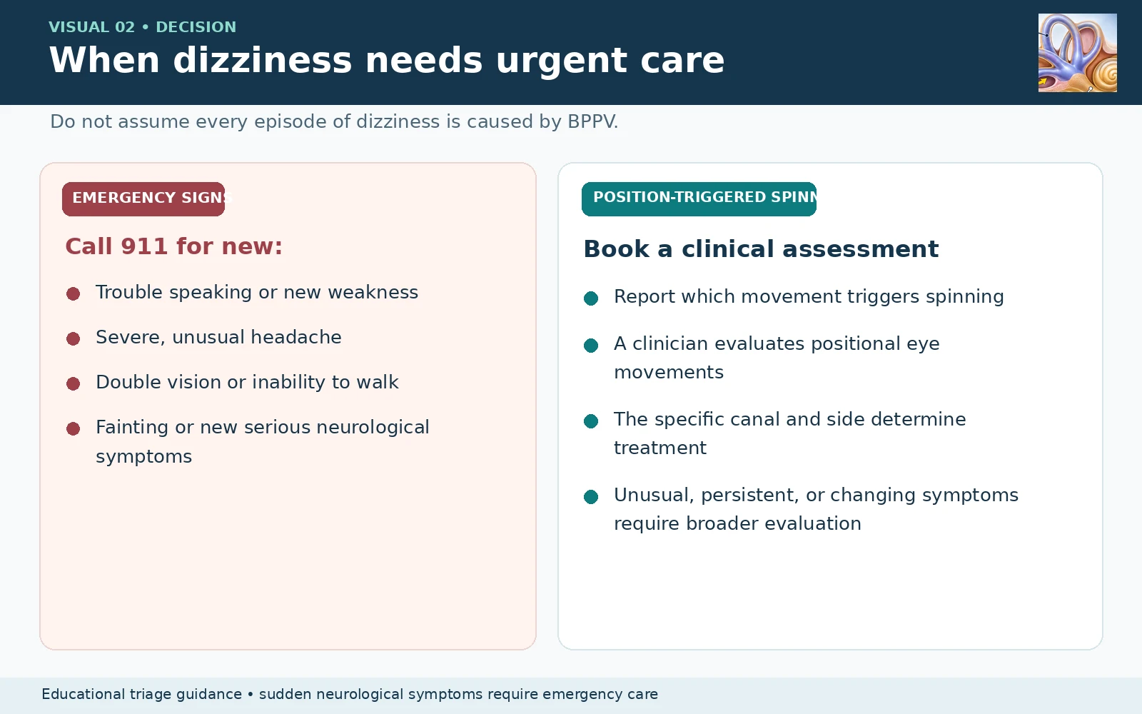
Call 911 immediately for sudden dizziness or vertigo with any possible stroke sign, including:
- Face drooping or new numbness/weakness, especially on one side
- Slurred speech, trouble speaking, or sudden confusion
- New double vision, vision loss, or other sudden major vision changes
- Sudden severe unexplained headache or neck pain
- New inability to stand or walk normally, severe loss of coordination, or unexplained collapse
- Chest pain, severe shortness of breath, or fainting with concerning symptoms
Do not drive yourself to the emergency department. Stroke can occasionally cause dizziness even without the classic face or arm signs.
Seek urgent medical assessment for sudden hearing loss; new, persistent, or severe vertigo that does not fit the usual brief positional pattern; repeated vomiting or inability to keep fluids down; or rapidly worsening symptoms. Sudden hearing loss needs same-day assessment, and continuous severe vertigo or neurological concerns may require the emergency department rather than routine urgent care.
What to expect after treatment
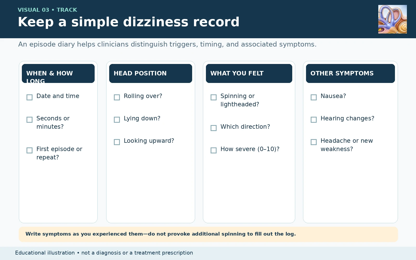
Many patients improve after one or several appropriately targeted repositioning sessions. Some experience temporary residual imbalance, and BPPV can recur. If symptoms persist or return, reassessment helps determine whether crystals remain displaced, a different canal is involved, or another condition needs attention. Patients should be careful with activities that increase fall risk while dizzy.
Questions about dizziness or positional vertigo?
ACE California Health Clinic provides chiropractic and acupuncture care in Santa Clara. Contact the clinic to discuss your symptoms, whether a vestibular evaluation is appropriate, and referral options when needed.
ACE California Health Clinic · 345 Saratoga Ave., Santa Clara, CA 95050
Phone: (408) 984-6000 · ACECaHealth.com
ACE California Health Clinic provides chiropractic and acupuncture care in Santa Clara. Contact the clinic to discuss your symptoms, whether a vestibular evaluation is appropriate, and referral options when needed.
ACE California Health Clinic · 345 Saratoga Ave., Santa Clara, CA 95050
Phone: (408) 984-6000 · ACECaHealth.com